The VExUS (Venous Excess Ultrasound) score is a clinical tool used to assess venous congestion in patients, often those with fluid overload or heart failure. This score combines measurements from several ultrasound modalities to evaluate the severity of congestion, which can influence management decisions in critical care settings. The grading ranges from 0 to 3, with Grade 3 indicating severe congestion.
Case Synopsis
Our patient is a 65-year-old female with a history of hypertension, coronary artery disease (CAD), and heart failure with an ejection fraction (EF) of 40%. She was admitted to the ICU with pneumonia and sepsis. After initial resuscitation with fluids, she developed renal impairment, raising concerns for fluid overload. To assess her volume status and guide diuretic therapy, a VExUS score was obtained.
Please review the above images and determine her VExUS score
VExUS grade is:
0%Grade 0
0%Grade 1
0%Grade 2
0%Grade 3
And now let's walk through the analysis of the case:
Step 1: Inferior Vena Cava (IVC) Diameter – Indicator of Central Venous Pressure

The first parameter in assessing venous congestion is the diameter of the inferior vena cava (IVC). A dilated IVC suggests elevated central venous pressure (CVP), which is often seen in fluid overload states.
In this case:
Observation: The IVC diameter is measured at 3.1 cm.
Interpretation: An IVC diameter greater than 2 cm is generally considered abnormally dilated. In this patient, a 3.1 cm measurement signifies a significant elevation in central venous pressure.
This finding points toward increased preload, which contributes to venous congestion and is a foundational component of a high VExUS score.
Step 2: Hepatic Vein Doppler – Reversal of the S Wave

Hepatic vein Doppler ultrasound assesses blood flow in the hepatic veins, which drain into the IVC. The normal waveform in hepatic vein Doppler should have a forward flow pattern, but reversal of the S wave indicates severe congestion.
In this case:
Observation: There is a clear reversal of the S wave, marked as severely abnormal.
Interpretation: The S wave reversal suggests retrograde blood flow due to elevated right atrial pressures and severe venous congestion. This finding is consistent with advanced right heart dysfunction or elevated right atrial pressure, as blood is impeded in returning to the heart.
This abnormality indicates severe hepatic venous congestion, which heavily contributes to a Grade 3 VExUS score.
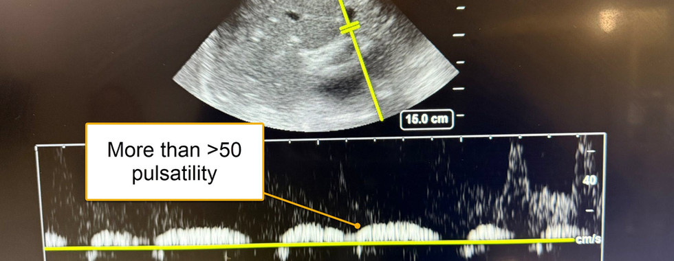
Step 3: Portal Vein Doppler – Pulsatility Index Greater than 50%

Portal vein Doppler can reveal pulsatility in the waveform, which normally should be minimal or absent in a healthy patient. Increased pulsatility here suggests backward transmission of venous pressure from the right side of the heart.
In this case:
Observation: The portal vein Doppler shows pulsatility greater than 50%.
Interpretation: A pulsatility index above 50% in the portal vein is considered severely abnormal and reflects high right atrial pressure that is transmitted to the portal system. This finding is indicative of severe portal venous congestion.
This is another significant marker of systemic congestion, reinforcing the high VExUS score.
Step 4: Renal Vein Doppler – Discontinuous Biphasic Flow

The renal vein Doppler assesses blood flow patterns in the renal veins, which can be altered in states of high venous pressure. Normally, the flow should be continuous, but discontinuity or biphasic flow suggests congestion.
In this case:
Observation: The renal vein Doppler demonstrates a discontinuous biphasic flow, which is moderately abnormal.
Interpretation: Discontinuous flow in the renal vein reflects moderate congestion affecting renal perfusion. It signifies that the venous congestion is now impacting renal blood flow, often associated with decreased kidney function due to elevated venous pressure.
This moderately abnormal finding further contributes to the elevated VExUS score, indicating renal venous congestion.
Conclusion (Grade 3 VExUS)
Combining these four findings, this patient was assigned a Grade 3 VExUS score, indicating severe systemic venous congestion. Here’s a summary of the abnormal findings leading to this score:
IVC Diameter greater than 2 cm (3.1 cm in this case) – suggests elevated central venous pressure.
Reversal of S Wave in hepatic vein Doppler – indicates severe hepatic venous congestion.
Portal Vein Pulsatility >50% – represents severe portal venous congestion.
Discontinuous Renal Vein Flow – indicates moderate renal venous congestion.
A Grade 3 VExUS score is associated with high degrees of venous congestion, which may necessitate therapeutic interventions such as fluid management or decongestive therapy. This scoring system allows healthcare providers to assess the extent of venous congestion and make informed management decisions, potentially improving outcomes for patients with severe fluid overload or heart failure.
References
Rola P, Miralles-Aguiar F, Argaiz E, Beaubien-Souligny W, Haycock K, Karimov T, Dinh VA, Spiegel R. Clinical applications of the venous excess ultrasound (VExUS) score: conceptual review and case series. Ultrasound J. 2021 Jun 19;13(1):32. doi: 10.1186/s13089-021-00232-8. PMID: 34146184; PMCID: PMC8214649.


Kaiser OTC benefits provide members with discounts on over-the-counter medications, vitamins, and health essentials, promoting better health management and cost-effective wellness solutions.
Obituaries near me help you find recent death notices, providing information about funeral services, memorials, and tributes for loved ones in your area.
is traveluro legit? Many users have had mixed experiences with the platform, so it's important to read reviews and verify deals before booking.
Introduction to NURS FPX 4060 Series Assessment
The NURS FPX 4060 series is a pivotal component of your nursing education, focusing on the integration of public health concepts into nursing practice. Covering NURS FPX 4060 Assessment 1 through NURS FPX 4060 Assessment 4, this series empowers you to address public health challenges effectively and contribute to the wellbeing of communities.