


The lung is anatomically divided into smaller units known as lobules, which are the smallest functional units of the lung visible to the naked eye. Each lobule is roughly hexagonal in shape and is bounded by connective tissue septa. Within these lobules, the lung's essential structures for gas exchange are contained, including terminal bronchioles, respiratory bronchioles, alveolar ducts, and alveoli. The terminal bronchiole, which is the smallest airway without alveoli, branches into several respiratory bronchioles, marking the transition where gas exchange begins. Blood supply to the lobules is via the pulmonary arteries and veins traveling through septa and branching along the bronchioles. Lymphatic vessels within the lobules help to maintain fluid balance and remove debris.


The "crazy paving" pattern on chest HRCT or standard CT refers to the appearance of ground-glass opacities with superimposed interlobular and intralobular septal thickening. This pattern is named for its resemblance to irregularly laid paving stones.

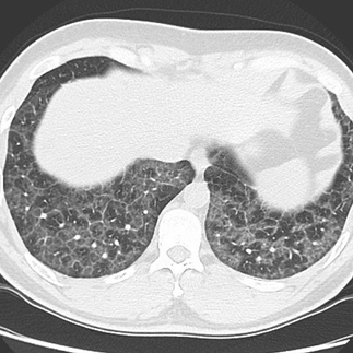


It is a non-specific finding that can be seen in a number of conditions affecting the lung parenchyma and interstitium, such as Pulmonary Alveolar Proteinosis (PAP), pulmonary edema, acute respiratory distress syndrome (ARDS), infectious pneumonias, interstitial lung diseases (ILD), lung hemorrhage, and lipoid pneumonia. The presence of this pattern prompts a detailed clinical and diagnostic workup to determine the underlying cause.