

23 years old male with mental status changes, tachycardia and suppressed TSH level.
Based on the above echocardiogram his ventricular function is:
0%Normal
0%Mildly decreaded
0%Moderately decreased
0%Severely decreased

23 years old male with mental status changes, tachycardia and suppressed TSH level.
Based on the above echocardiogram his ventricular function is:
0%Normal
0%Mildly decreaded
0%Moderately decreased
0%Severely decreased
Post cardiac arrest, CPR 15 min , ROSC achieved
VExUS for evaluation of venous congestion in a patient with mixed cardiogenic and distributive shock, acute respiratory failure, bibasilar infiltrates, and acute kidney injury.
Step 1: IVC diameter is 2.5 cm

Step 2: hepatic vein doppler assessment showing reversal of the systolic wave.

Step 3: Portal vein doppler assessment showing more than 50% variability
Very beautiful demonstration
It helps to suggest offloading rather than fluid resuscitation
It requires stady hands and minimal pt movement (abd wise) in order to catch the image in PW mode
If my memory serves me right, there were only limited data from multiple case report
No strong data to suggest outcome
Yet a useful tool to add on for the management
If we have research, i think mortality outcome, Cr trend, O2 requirements, vasopressors doses would be excellent markers for monitoring
Thank you Dr Mazen for your amazing continuous contribution to enriching our knowledge
Bedside echocardiography showing severely depressed LV function!
Severe mitral valve regurgitation:
The mitral valve is a leaflets valve that separates the left atrium from the left ventricle. The valve opens to allow blood flow from the atrium to the ventricle during diastole, and closes to prevent backflow from the ventricle to the atrium during systole. MR occurs when the mitral valve does not close properly, resulting in leakage of blood back into the left atrium during systole. This results in volume overload of the left atrium, which can lead to left atrial enlargement and, ultimately, left ventricular dysfunction.
Transthoracic echocardiography (TTE) is typically used as the initial screening test, as it is widely available, relatively inexpensive, and can be performed quickly at the bedside. TTE allows for assessment of heart size, valvular anatomy, and valvular function. Doppler echocardiography can be used to quantify MR by measuring gradient across the valve, by assessing regurgitant volume, or by measuring…

Tricuspid annular plane systolic excursion (TAPSE) is a parameter of global right ventricular function. TAPSE is the displacement of the tricuspid annulus in systole, normally about 18-22 mm. It is measured by M-mode echocardiography with the curser over the lateral aspect of the tricuspid annulus. Measure the distance of the maximal longitudinal displacement and obtain the average of three consecutive cardiac cycles. TAPSE correlates well with other measures of right ventricular function such as RV ejection fraction. TAPSE can be used to diagnose pulmonary embolism, as it is significantly reduced in patients with PE. TAPSE can also be used to assess response to therapies for PE and determine prognosis. Place the M-mode cursor over the lateral aspect of the tricuspid annulus. Measure the distance of the maximal longitudinal displacement. Calculate the average of three consecutive cardiac cycles
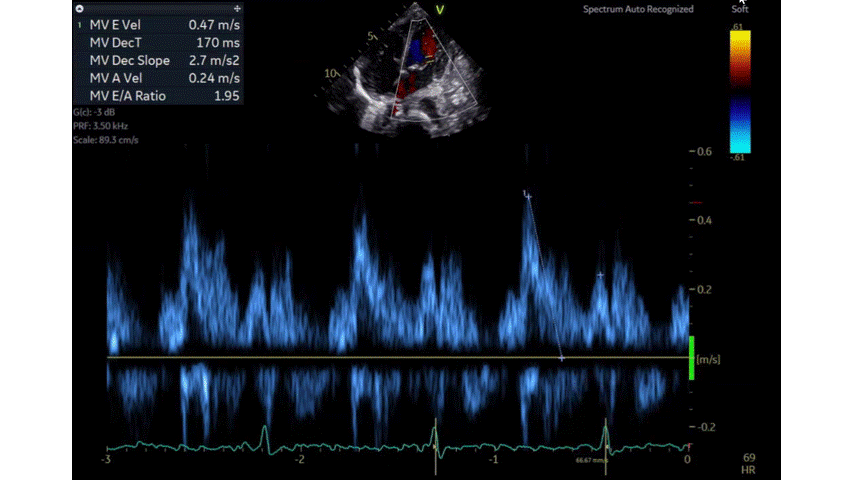
Diastolic heart function refers to the filling of the ventricles with blood during diastole, or the relaxation phase of the heart. Diastolic dysfunction occurs when the ventricles do not fill properly or evenly. Estimating diastolic heart function is important in order to diagnose and treat this condition. Echocardiography is a non-invasive imaging technique that can be used to estimate diastolic function. Doppler velocity echocardiography is a specific type of echocardiography that uses sound waves to measure blood flow velocity. E wave represents the peak velocity blood flow from left ventricular relaxation in early diastole (correspond to the V wave on CVP waveform), and the A wave represents the peak velocity flow in late diastole caused by atrial contraction (correspond to the A wave on CVP waveform). The E wave is normally higher than the A wave with a normal E/A ratio of ≥0.8. E/A ratio is a marker of the…
Echo was done by on of the IM residents in the ICU at the bedside, what do you think of the cardiac function?
Limited echo due the technique but shows severely depressed biventricular function! Bedside echocardiogram by the intensivist may add valuable information that can help in the management of the patient.
 Creator
I think the proper is flipped
The large akentic chamber in the left has thick wall similar to LV