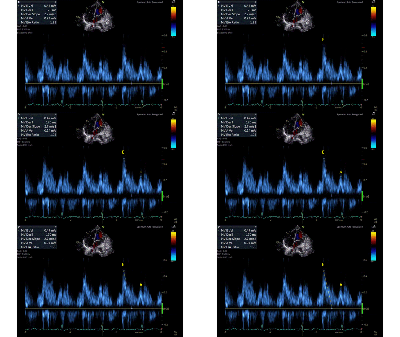
Diastolic heart function refers to the filling of the ventricles with blood during diastole, or the relaxation phase of the heart. Diastolic dysfunction occurs when the ventricles do not fill properly or evenly. Estimating diastolic heart function is important in order to diagnose and treat this condition. Echocardiography is a non-invasive imaging technique that can be used to estimate diastolic function. Doppler velocity echocardiography is a specific type of echocardiography that uses sound waves to measure blood flow velocity. E wave represents the peak velocity blood flow from left ventricular relaxation in early diastole (correspond to the V wave on CVP waveform), and the A wave represents the peak velocity flow in late diastole caused by atrial contraction (correspond to the A wave on CVP waveform). The E wave is normally higher than the A wave with a normal E/A ratio of ≥0.8. E/A ratio is a marker of the diastolic function of the left ventricle when it is less than 0.8. A psudonormal E/A ratio can be seen and tissue doppler septal annulus (e`) would be needed to determine diastolic dysfunction.
In the above doppler image, the MV E Velocity is 0.47 m/s and the MV A Velocity is 0.24 m/s with a normal E/A ratio of 1.95. The yellow line is the descending slope of the E wave.


